RESOURCES

Where research scientists come to find up to date resources on antibody discovery against challenging membrane protein targets.

Category: Specificity Insights

The Consequences of Off-Target Binding Are Real. So Are the Solutions.

In this article:

This article is part 6 of a series about specificity testing.

Summary

Off-target binding doesn’t always cause harm—but when it goes undetected, the consequences can be serious. Published case studies document off-target interactions that caused adverse events in clinical trials, halted drug development programs, and may explain the known safety profiles of approved therapeutics. These cases also demonstrate that when off-targets are identified early, solutions exist: engineering fixes, informed go/no-go decisions, and cleaner IND filings.

Can off-target binding cause adverse events?

In the previous article, we looked at how common off-target binding is among biotherapeutic drug candidates. Here, we look at what undetected off-target binding can mean in practice.

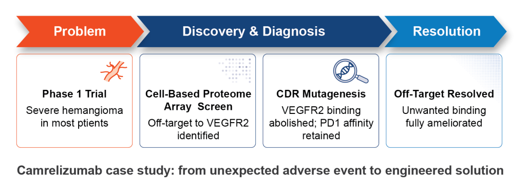
One of the most well-documented examples of off-target-driven toxicity involves camrelizumab, an anti-PD1 monoclonal antibody developed for cancer immunotherapy. During Phase 1 clinical trials, a majority of treated patients developed severe capillary hemangioma. These abnormal blood vessel growths had no obvious connection to PD1, the antibody’s intended target (Mo et al., 2018).

Retrospective screening of camrelizumab using a cell-based protein array identified the source of the problem: the antibody was also binding VEGFR2, a receptor with a central role in blood vessel formation (Finlay et al., 2019). And the binding wasn’t passive. Camrelizumab acted as an agonist, actively stimulating VEGFR2 to promote vascular neogenesis, which, in vivo, leads to hemangioma.

The causal link was confirmed when patients treated with a VEGFR2 antagonist saw the hemangioma effects resolved (Li et al., 2019). Equally significant: CDR mutagenesis abolished binding to VEGFR2 while preserving high affinity for PD1 (Finlay et al., 2019). Had it been found earlier, the off-target binding could have been engineered away prior to trials in patients.

Timeline of the camrelizumab story, from problem to diagnosis, CDR engineering, and resolution.

What does MPA screening of clinical-stage MAbs reveal?

Looking more broadly, Membrane Proteome Array (MPA) screening of clinical-stage monoclonal antibodies has identified several cases where off-target binding may explain known adverse events (Norden et al., 2024). Among them:

  • A clinical-stage MAb armed with a toxic payload was found to bind an off-target protein with widespread tissue expression—a combination predicted to cause significant toxicity.
  • A MAb that was withdrawn from clinical trials due to severe patient adverse events was found to bind an unrelated membrane protein even more strongly than its intended target.

In these cases, the relationship between the off-target interaction and the observed clinical outcome is not fully established. What is clear is that the off-target binding existed, went undetected by prior specificity assessments, and was later identified by MPA screening.

Can secreted protein off-targets pose safety and efficacy risks?

Membrane proteins are the primary focus for specificity testing because off-target binding to them carries the highest safety risk, but off-target binding to secreted proteins can also carry consequences. While off-target binding to secreted proteins is less likely to cause cell death or direct disruption of cell biology, toxicity has still been reported.

In one case, development of ABT-736, an anti-beta-amyloid antibody, was discontinued after it caused severe toxicity in cynomolgus monkeys. This toxicity was later traced to off-target binding to platelet factor 4, a secreted plasma protein (Loberg et al., 2021).

Beyond direct toxicity, off-target binding to abundant circulating proteins can also affect pharmacokinetics and dosing (Bumbaca et al., 2011).

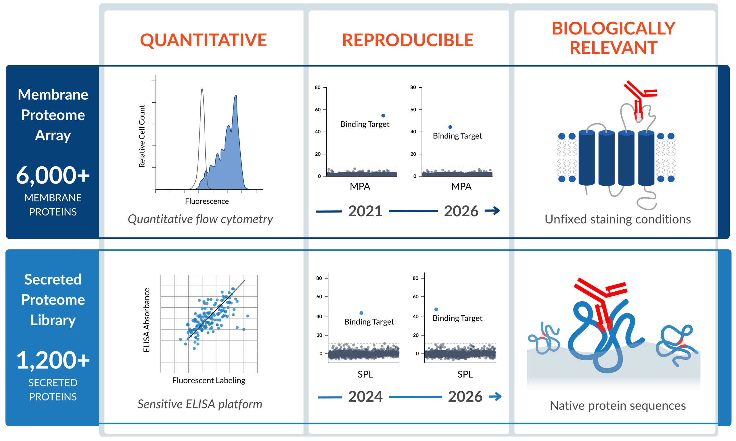
To identify potential secreted off-targets, and to provide a comprehensive picture of your molecule’s interactions with the human body, Integral Molecular offers screening against over 1,200 soluble proteins on the Secreted Proteome Library (SPL). We’ll share more about the SPL in a future article.

Do off-targets always cause adverse events?

The camrelizumab story could have gone very differently if the VEGFR2 interaction had been identified preclinically. CDR mutagenesis, the same approach that was used retrospectively, could have been applied before patients were enrolled.

This is what earlier, more comprehensive specificity testing enables: not just detection of off-targets, but the opportunity to act on that information while options still exist. For candidates without off-target interactions, MPA results can be included directly in IND filings. For candidates with off-target interactions, the MPA identifies the specific protein involved, enabling a focused investigation rather than a search for an unknown cause. While off-target binding does not always lead to adverse events, it does always warrant investigation.

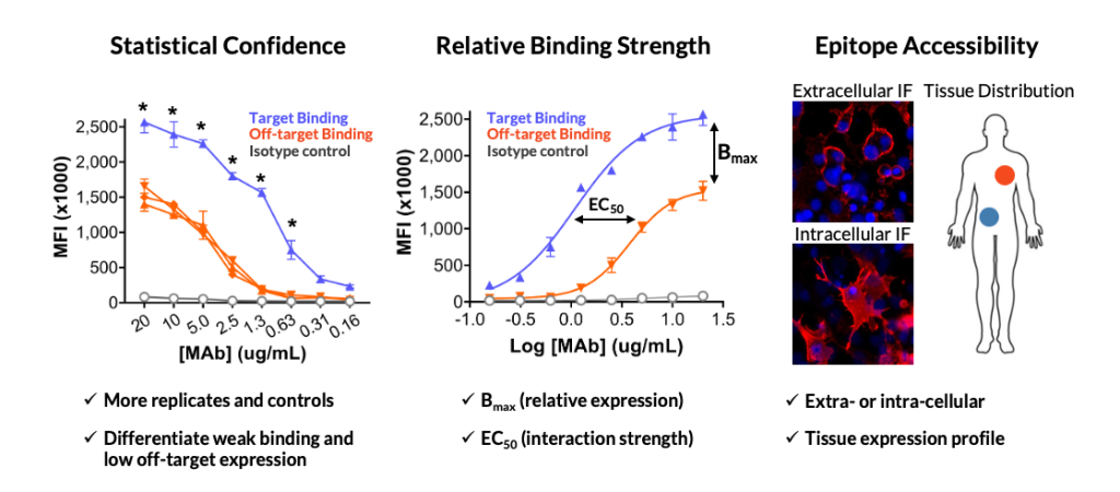
Any investigation into an off-target should consider factors including the relative strength of the off-target interaction compared to on-target binding, the epitope location and accessibility of the off-target protein, and the therapeutic mechanism of action. Integral Molecular’s Enhanced Binding Analysis service provides the quantitative data needed to begin that evaluation.

Understanding these factors will help you understand the associated risk and arm you with the information you need to make decisions. For example, a low-affinity interaction with an intracellular epitope presents a very different risk profile than strong binding to a widely expressed cell-surface protein—particularly for cell-killing modalities like ADCs or CAR-T therapies.

Enhanced Binding Analysis performs a statistical assessment, measures relative binding strength, and evaluates epitope accessibility.
Enhanced Binding Analysis provides information about target binding, accessibility, and potential for therapeutic toxicity.

Looking ahead

In the next article, we’ll cover how the MPA library was designed, and what it means for a specificity testing tool to be truly comprehensive.

1 in 3 Antibody Drug Candidates Bind Off-Targets. Here’s What That Means.

In this article:

This article is part 5 of a series about specificity testing. Be sure to read part 3, about the limitations of tissue cross-reactivity studies. 

Summary

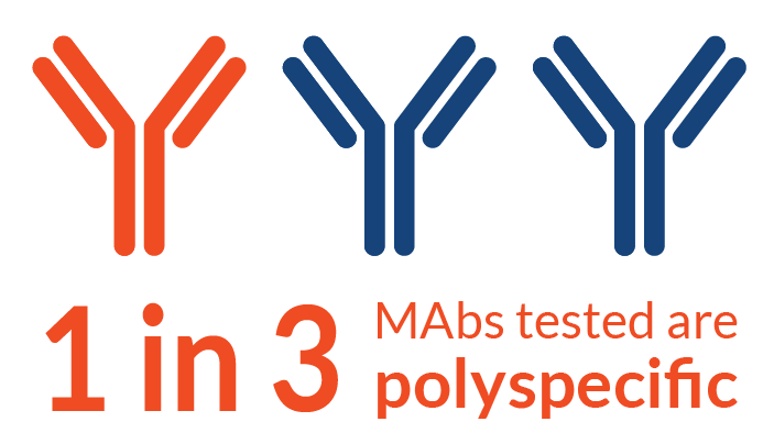
Off-target binding in antibody therapeutics is far more common than most people expect. Our analysis of hundreds of antibody-based drug candidates found that roughly 1 in 3 lead candidates display polyspecific off-target binding. And the problem persists as drugs advance through development. Even among FDA-approved MAbs, about 15% show off-target binding. Side-by-side comparisons of MPA and tissue cross-reactivity (TCR) data reveal that many of these off-targets went undetected by traditional methods. Spotting these interactions early, and identifying the specific off-target proteins, is the first step toward addressing them.

How common is off-target binding, really?

In an earlier article, we mentioned that 1 in 3 antibody lead candidates has off-target binding. Here, we’ll dig into that number.

To quantify the prevalence of polyspecificity across the industry, we conducted a retrospective analysis of all antibody-based therapeutics submitted by customers for specificity testing on the Membrane Proteome Array (MPA) over a defined time period. We analyzed 254 samples in total, including MAbs, scFv-Fcs, and VHH-Fcs. These samples primarily represent lead candidates at biopharmaceutical companies throughout the industry. To be included in the analysis, each sample had to have successfully completed all three steps of the MPA process: Assay Setup, MPA Screen, and Validation (Norden et al., 2024).

The results were striking:

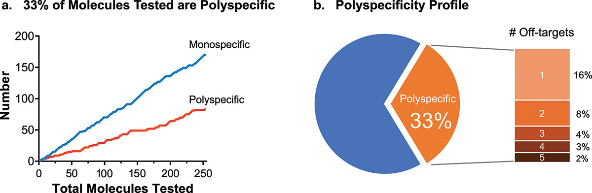
  • 83 of 254 samples (32.7%) displayed polyspecific off-target binding.
  • Among the polyspecific antibodies, about half had a single off-target, while the rest had two or more.
  • Off-target binding was almost always to completely unpredictable membrane proteins with no significant sequence homology to the intended target.

Graph of total molecules tested over time x number of monospecific and polyspecific molecules reveals that 33% of test articles are polyspecific. Pie graph shows that of this 33%, 16% have one off-target, 8% have 2, and 9% have 3 or more.

33% of 254 antibody-based lead candidates screened on the MPA demonstrated validated off-target binding. Data from Norden et al., 2024.

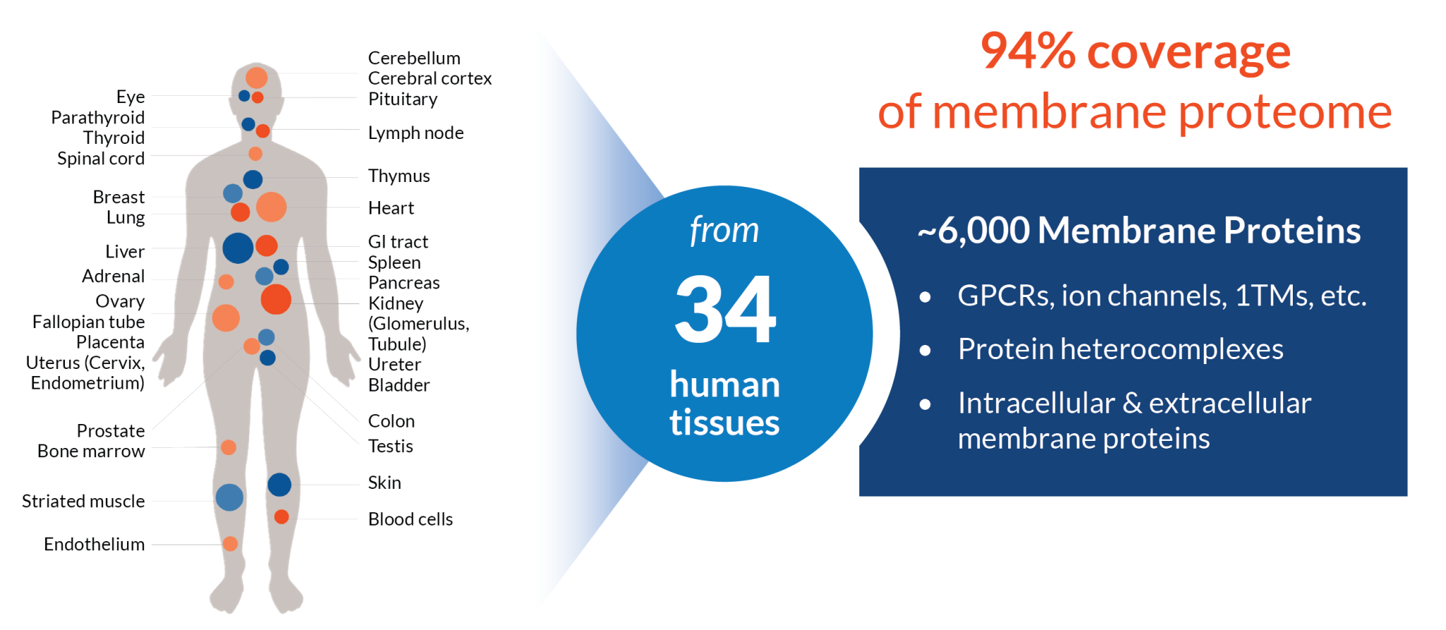
The 33% figure reflects true, confirmed off-target binding interactions. Off-target binding was detected during MPA screening on ~6,000 native membrane proteins, then validated by antibody titration studies to confirm each hit. Note that these results include only CDR-mediated interactions. Non-CDR-mediated interactions, such as binding to Fc receptors or lectins, would push the off-target rate even higher if counted.

Perhaps most importantly, the off-targets were almost never related to the intended targets. In part, that’s because most MAbs screened on the MPA have already been tested against members of the same protein family. The study results represent genuinely unexpected interactions that conventional approaches would miss entirely.

The findings support our recommendation to conduct specificity testing using cell-based protein arrays early in drug development, ideally during lead selection, when potential toxicity issues can be identified and addressed with relatively little impact on a drug program.

Does polyspecificity persist into late-stage development?

Given the high off-target rate among lead candidates, we wanted to know whether polyspecificity persists among MAbs that have gone into humans. To find out, we produced biosimilars of 83 clinical-stage, FDA-approved, and withdrawn MAbs and screened them on the Membrane Proteome Array (MPA).

The answer is yes: polyspecificity persists at every stage of clinical development.

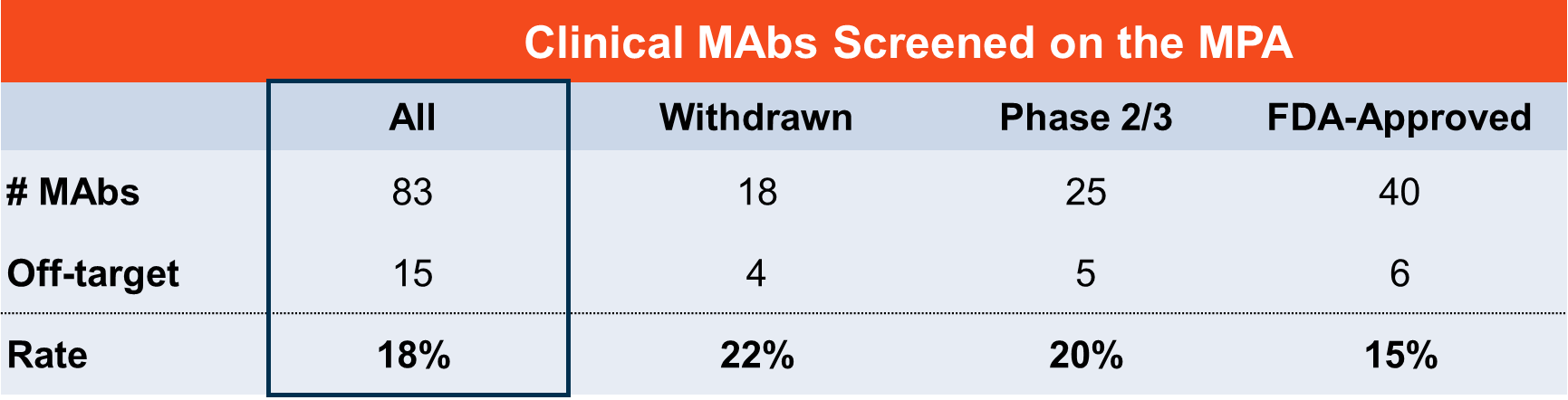
  • 18.1% of clinical MAbs overall showed off-target binding.
  • Off-target rates were slightly higher for withdrawn MAbs (22.2%) and those in Phase 2/3 (20.0%) compared to approved MAbs (15.0%).

Clinical MAbs screened on the MPA. Table with number of antibodies, number of off-targets, and off-target rate by status: withdrawn, phase 2/3, FDA-approved, and total

Off-target binding was found in MAbs at all stages of clinical development, with higher rates among withdrawn and clinical-stage MAbs than approved ones. Based on Norden et al., 2024.

The lower rate among clinical-stage MAbs compared to lead candidates (18% vs. 33%) suggests that polyspecificity contributes to drug attrition; candidates with off-target binding appear to drop out of the pipeline at higher rates. But an off-target binding rate of 15% in FDA-approved drugs makes clear that current screening methods are not catching everything.

An independent analysis published in 2026 reached a similar conclusion. Dai et al. used a different platform to screen 174 FDA-approved and clinical-stage MAbs against 6,172 human extracellular proteins, finding that 28% had at least one off-target. The Dai et al. and Norden et al. studies used different technologies and somewhat different protein sets, but both point to the same conclusion: off-target binding among clinical antibodies is not rare, and it is not an artifact of any single platform.

Why can’t you predict off-target binding from sequence analysis alone?

One of the most consistent findings across our Membrane Proteome Array (MPA) data is that off-target binding is almost never to a related protein. So why is it happening?

Three mechanisms have been identified:

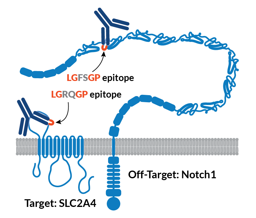
Molecular mimicry is likely the most common. Critical epitope residues in the intended target are mirrored, by chance, in a completely unrelated protein. In one well-documented example, a MAb we isolated against the glucose transporter SLC2A4 (GLUT4) also bound to Notch1, a signaling protein with less than 7% sequence identity and no structural similarity to GLUT4. Epitope mapping traced the cross-reactivity to a shared LGXXGP motif present in both proteins: one in a loop on GLUT4, the other in a disulfide-constrained loop on Notch1 (Tucker et al., 2018).

In an example of molecular mimicry, the target and a completely unrelated off-target share an LGXXGP epitope

Molecular mimicry explains how a MAb against SLC2A4 (GLUT4) also bound Notch1—despite less than 7% sequence identity. Epitope mapping revealed that both proteins share an LGXXGP epitope motif. Graphic based on Norden et al., 2024, Fig. 6.

CDR plasticity is a second mechanism, in which conformational flexibility in the antibody’s complementarity-determining regions (CDRs) allows the paratope to adapt to more than one antigen.
Differential CDR engagement is a third possibility: off-target binding may occur through entirely different CDR residues than those used to bind the primary target.

The practical implication is the same regardless of mechanism: off-target binding cannot be reliably predicted from sequence or structural analysis. Proteome-wide empirical screening is the only reliable way to detect it.

What is TCR missing?

As discussed in an earlier article, tissue cross-reactivity (TCR) studies have significant limitations. Our own data provides direct evidence of one of the most consequential: TCR is missing off-target interactions that the Membrane Proteome Array (MPA) detects.

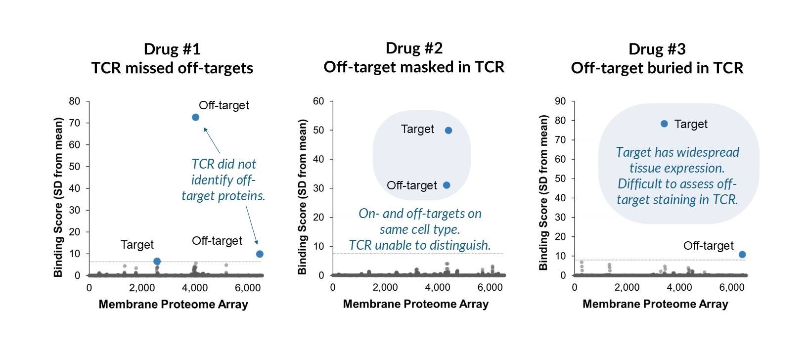
To compare the two approaches, we reproduced FDA-approved MAbs and screened them on the MPA, then compared our results to TCR data from the corresponding biologics license applications (BLAs). In several cases, the MPA identified off-target binding that was not mentioned in the TCR summaries.

MPA data plots for the three case studies described in the text, where TCR missed off-targets, off-targets were masked in TCR, and an off-target was buried amid TCR staining.

In three side-by-side comparisons, MPA screening of biosimilars of FDA-approved MAbs identified off-targets that were not detected or reported in available TCR data from BLA applications. Dotted line represents 3 SD above calculated background. Adapted from Norden et al., 2024.

Three examples illustrate the pattern:

  • Case Study 1. This antibody targets a plasma membrane protein expressed on lymphocytes. The MPA correctly identified the intended target—and also detected two off-target membrane proteins that bound even more strongly than the target. The BLA summary indicated TCR staining consistent with known target expression on lymphocytes, with no off-target binding reported.
  • Case Study 2. This antibody targets a membrane protein expressed on the plasma membrane and intracellularly in myeloid cells. The MPA identified the target and an additional off-target expressed on the same cell type. The BLA summary noted staining consistent with known target expression and did not mention off-target binding. When the target and off-target are co-expressed on the same cell, tissue-based methods cannot distinguish between them.
  • Case Study 3. This antibody targets a membrane protein with low, widespread expression across most normal tissues that is upregulated in certain disease states. The MPA identified the target and an off-target membrane protein. The BLA summary reported primarily cytoplasmic staining across many tissues, with no significant off-target concerns—likely because identifying an off-target signal against a background of widespread positive staining is extremely difficult using TCR.

In none of these cases were the off-targets members of the same protein family as the intended target. These represent genuinely unexpected interactions that TCR was not designed to detect.

Looking ahead

The data clearly show that off-target binding is a widespread problem that traditional methods are failing to catch. But what actually happens when those interactions go undetected—and make it into the clinic? In the next article, we examine case studies where undetected off-target binding led to serious adverse events in patients, and what those examples tell us about the need for better specificity testing.

How Is the MPA Being Qualified by the FDA?

In this article: 

This article is part of a series about specificity testing. Be sure to read part 3, about the limitations of tissue cross-reactivity studies. 

Summary 

Tissue cross-reactivity (TCR) studies have served as the standard for specificity testing since the 1980s, but their limitations—inability to identify specific proteins, subjective interpretation, poor clinical correlation—have long been recognized. The Membrane Proteome Array (MPA) was designed to address these gaps with objective, quantitative data that identifies specific off-target proteins. In 2021, we began working with the FDA to qualify the MPA as a Drug Development Tool through the ISTAND program. While the FDA already accepts MPA data, this qualification process validates the scientific rigor behind the platform and helps formalize the shift toward more predictive, human-relevant specificity testing methods.

Why we built a better specificity testing tool

As discussed in the previous article, tissue cross-reactivity (TCR) studies leave significant gaps in our understanding of therapeutic specificity. Most critically, they can’t tell you which protein a therapeutic is binding to—only where unexpected staining appears in tissues. That makes it nearly impossible to assess the actual safety risk or design appropriate follow-up studies. 

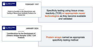
Drug developers and regulators have long recognized the need for better tools. The FDA stated as far back as 1997 that “appropriate newer technologies should be employed as they become available and validated.” More recently, their 2024 CAR-T guidance specifically named “protein arrays” as an acceptable alternative to TCR. 

The Membrane Proteome Array (MPA) was designed to fill the gaps left by TCR. And the shift in the regulatory landscape has already begun: MPA data has already been accepted in over 100 IND applications. Proper specificity assessment early in development contributes to better candidate selection, more efficient regulatory review, safer clinical trials, and ultimately better therapeutics for patients. 

What advantages does the MPA have over conventional methods?

The MPA offers several key advantages over conventional specificity testing methods: 

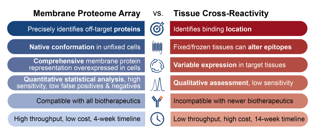
      • Identifies specific proteins. When the MPA detects off-target binding, it tells you exactly which proteins are involved. That enables focused investigation into potential safety issues and informed decisions about whether to proceed with development. TCR studies, by contrast, can only show you tissue locations. 
      • Uses native protein conformations. Each protein is expressed in its natural state within whole eukaryotic cells, with proper folding, post-translational modifications, and membrane environment. TCR tissue processing can alter protein structures, potentially causing false positives or negatives.  
      • Eliminates donor variability. The MPA uses established cell lines with consistent handling protocols, so every protein is fully expressed and available for testing. TCR’s reliance on tissue samples from three individual donors means protein expression varies unpredictably. 
      • Provides objective, quantitative data. The MPA uses flow cytometry to generate measurements that can be statistically analyzed and compared across studies. This eliminates the subjectivity inherent in TCR, where pathologists score immunohistochemistry staining patterns.  

Additionally, MPA data supports regulatory submissions directly. MPA results provide the precise, quantitative data needed for IND applications. For candidates without off-targets, the specificity is demonstrated clearly. For those with off-targets, you have the molecular detail needed to design appropriate follow-up studies. 

These advantages made the MPA an ideal candidate for FDA qualification as a Drug Development Tool—a formal recognition that would help accelerate the broader shift toward more predictive specificity testing methods.

Specificity testing on the Membrane Proteome Array offers many advantages over tissue cross-reactivity studies.

What is the ISTAND qualification process?

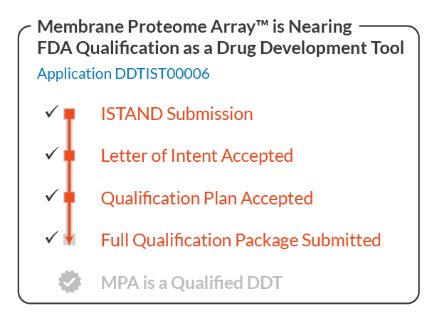
In May 2021, Integral Molecular submitted a Letter of Intent to the FDA’s ISTAND program to qualify the MPA as a Drug Development Tool. ISTAND (Innovative Science and Technology Approaches for New Drugs) provides a pathway for qualifying novel methods that can improve drug development and regulatory review. 

The FDA accepted the MPA into ISTAND in July 2022, making it the first tool ever accepted into the program. This milestone reflected both the platform’s scientific merit and the FDA’s recognition that better specificity testing methods are needed. 

The qualification process involves several stages: 

      • Letter of Intent (LOI) - Describes the tool, its intended use, and preliminary data supporting its utility. Accepted July 2022. 
      • Qualification Plan (QP) - Outlines the validation studies, performance characteristics, and regulatory strategy. Submitted August 2023, accepted January 2025. 
      • Full Qualification Package (FQP) - Provides comprehensive validation data and evidence supporting the tool’s use in regulatory submissions. Submitted Q4  2025. 

Throughout this process, the FDA has provided feedback and suggestions for enhancements. Their input has helped transform an already strong platform into one that’s even more robust, reproducible, scalable, and well-documented. 

The MPA is nearing FDA qualification as a drug development tool. Qualification is a rigorous, multi-step process, for which we have submitted all required documents.

What makes the MPA’s qualification significant?

While the FDA already accepts MPA data, qualification will streamline the regulatory review process. Once the FDA qualifies the MPA, any drug developer can use it in their IND applications “with confidence that the FDA will accept the data” (Roadmap to Reducing Animal Testing in Preclinical Safety Studies). 

The MPA will likely represent several firsts in the New Approach Methodology (NAM) and Drug Development Tool landscape: 

      • The only NAM specificity test with published validation data 
      • The first qualified NAM for specificity testing 
      • On track to be the first NAM qualified through the ISTAND process 
      • On track to be the first NAM qualified as a DDT by the FDA 

For organizations considering qualifying their own drug development tools through ISTAND, we highly recommend it. The experience has been invaluable, and the FDA’s input has genuinely enhanced the platform. 

Looking ahead 

In the coming articles, we’ll share more details about how the MPA works, including the proteins represented in the library, the screening process, and the quality systems that ensure reliable results. We’ll also discuss how MPA data compares to TCR studies and share insights into minimizing false positives and false negatives. 

Are Tissue Cross-Reactivity Studies Sufficient for Biotherapeutic Specificity Testing?

In this article:

This article is part 3 of a series about specificity testing. Be sure to read part 2, about the available tools and when to use them.

Summary 

The short answer is no, tissue cross-reactivity (TCR) studies are not sufficient for biotherapeutic specificity testing. Even though TCR is widely used and has long been recommended by the FDA, it has a long and much-discussed list of limitations. Importantly, TCR results correlate poorly with clinical outcomes, and toxicologists don’t often use TCR data to drive drug development decisions. There is a clear need for a shift to newer specificity testing approaches that are quantitative, objective, validated, and better correlated with patient safety outcomes.

Why are conventional specificity testing methods insufficient?

As described in the previous article in this series, What tools are available for specificity testing during drug development?, tissue cross-reactivity (TCR) studies were the first specificity testing method to be required for biotherapeutic IND submissions beginning in the 1980s. TCR was the best specificity testing tool available at the time, and it quickly became the standard. But as most people familiar with TCR will tell you, the technology has inherent limitations. Importantly, its results correlate poorly with patient safety outcomes.

This article breaks down those limitations, presents some information about how toxicologists use (or don’t use) TCR data, and points to several resources where you can learn more about these topics.

TCR study
TCR is in vitro assay that uses immunohistochemistry (IHC) to reveal antibody binding across a panel of human tissue samples from three individual donors. Results are interpreted by a pathologist, who looks for unexpected off-target binding and previously unknown sites of on-target binding.

What does the literature say about the limitations of TCR studies?

Several published reviews (Cunningham et al., 2021; Li et al., 2020; MacLachlan et al., 2021 ) and case studies (Brennan et al., 2018; Leach et al., 2010) walk through the limitations of using TCR studies for predicting in vivo toxicity and safety.

The following list, pulled from the resources above and our own publications and experiences, summarizes TCR’s most concerning limitations.

  1. TCR cannot identify target proteins. The primary limitation of TCR studies is that they can identify only binding locations; they cannot reveal the identity of specific target or off-target proteins. That means if TCR reveals unexpected binding patterns, the options for follow-up studies to understand the nature of that binding are severely limited. Moreover, on-target tissue staining may provide false reassurance when a target and off-target are expressed within the same tissue or when the off-target is expressed at low levels.
  2. In vivo protein expression is highly variable. Protein expression levels vary within and between tissues, between individuals, and over time—and these levels are difficult to quantify. With TCR, there’s no way to determine whether lack of staining is due to lack of antibody-protein interaction or lack of protein expression.
  3. Processing alters protein conformations. Most TCR tissue samples are fixed or frozen, placed onto glass slides, and processed for staining. These steps can alter proteins’ conformations, potentially leading to false positives and false negatives.
  4. TCR has high background staining levels. Native Fc receptors and endogenous IgG present throughout human tissues frequently cause high background binding, complicating results interpretation.
  5. Scoring is subjective. All TCR results are based on qualitative observations. Trained pathologists score IHC staining results based on their observations and interpretations.
  6. Turnaround is slow. Optimizing the staining protocol and completing TCR studies typically takes 12 weeks or more.
  7. Secreted proteins are excluded. Secreted proteins can be a source of off-target binding, but they are washed off the tissue samples during processing. Thus, TCR cannot reveal target or off-target binding to secreted proteins.
  8. Some protein variations remain untested. Tissue samples from three donors are unlikely to include all possible protein variations present in the general population:
    • Heterocomplex formation and multimer arrangements are often transient or disease specific.
    • Many targets can be expressed as several possible isoforms and have isoform- or disease-specific cellular locations.
    • Post-translational modifications are variable and they can be permanent or transient.
  9. TCR studies are not quantitative. With no quantification, TCR studies cannot be statistically analyzed or tracked for quality control.
  10. TCR studies have never been validated. There are no published analyses of measures such as reproducibility, variability, false-negatives, false-positives, and sensitivity.
  11. TCR studies have never been qualified by the FDA. Although TCR studies are accepted, TCR studies have never formally been qualified by the FDA, who would review the available validation data if it existed.

The long list of limitations shows that while TCR can provide useful information about biotherapeutic binding locations, other tools are needed to better detect and understand off-target binders and predict patient safety outcomes.

Do toxicologists trust TCR for determining specificity?

Given its long list of limitations, it shouldn’t be too surprising that toxicologists don’t trust TCR data. Perhaps the most telling indication of this viewpoint is a set of survey results that captures just how little influence TCR studies have on drug development decisions (MacLachlan et al., 2021). In this survey, industry experts, mostly biotechnology and pharmaceutical toxicologists, answered a series of questions about how they perceive the utility and value of TCR studies.

The results indicate that the vast majority of toxicologists believe TCR results are not predictive of in vivo toxicity, and that TCR results are not actually used in practice to make critical decisions. We encourage you to read the paper.

TCR survey

Cell-based protein arrays fill the gaps

TCR studies leave large information—and trust—gaps. These gaps could be filled by an objective, quantitative, and consistent approach that identifies target proteins and enables statistical analysis between studies.

Fortunately, such tools already exist, and they are increasingly being adopted. Cell-based protein arrays, such as the Membrane Proteome Array (MPA), provide essential information beyond what TCR can deliver.

As this series continues, we’ll share more about cell-based protein arrays and the evolving specificity testing landscape, including what it looks like to qualify a New Approach Methodology (NAM) with the FDA, how MPA and TCR compare in performance, and methods for minimizing false positives and false negatives.

Looking ahead 

In the next article, we’ll dig into how the MPA addresses TCR limitations, and how it is being qualified through the FDA’s ISTAND program.

What Tools Are Available for Specificity Testing During Drug Development?

In this article:

This article is part 2 of a series about specificity testing. Be sure to read part 1, an introduction to the series. 

Summary 

Even though monoclonal antibodies are prized for their specificity, 1 in 3 lead candidates exhibit off-target binding. Because off-target interactions are so common and their safety consequences so large, we need accurate methods for identifying them. Specificity testing during preclinical development typically involves one or both of two technologies: tissue cross-reactivity (TCR) studies, which have been in use since the 1980s, and cell-based protein arrays, which have emerged in the last decade or so. Other methods, including tissue microarrays and spotted protein microarrays, have limitations that make them inappropriate for preclinical safety testing. While early FDA guidance focused on TCR, recent documents have named protein arrays as an appropriate alternative. 

Why is specificity testing important?

Monoclonal antibodies (Mabs) are prized for their exquisite specificity—especially compared to their small-molecule counterparts. Yet a study of therapeutic MAbs in development and on the market showed that 1 in 3 lead candidates exhibited off-target binding (Norden et al., 2024). 

Given the potential consequences of off-target interactions—on patient safety, drug efficacy, pharmacokinetics—it’s essential to have accurate methods for identifying them during preclinical development. That way, we can advance only the safest, most-selective drugs to the clinic. 

In this article, we’ll break down the methods that are currently available for preclinical specificity testing and describe how FDA guidance in this realm has changed over time. 

What is off-target binding?

Off-target binding, also known as cross-reactivity or polyspecificity, is CDR-mediated binding to one or more proteins other than the intended target. Sometimes the off-target is a member of the same protein family, but most often it’s a completely unrelated protein and thus extremely difficult to predict.  

That’s in contrast to polyreactivity, which refers to a more general type of antibody stickiness—an antibody developability challenge in its own right. There are well-known methods for detecting polyreactivity, though we won’t discuss them here.  

For a deeper discussion of polyreactivity and polyspecificity, see Cunningham et al., 2021. 

What off-target specificity testing methods are currently available?

While the FDA has yet to qualify any tools for evaluating biotherapeutic specificity, several technologies exist for this purpose. Specificity testing during preclinical development typically involves tissue cross-reactivity studies, cell-based protein arrays, or both. Other methods, including tissue microarrays and spotted protein microarrays, have limitations that make them inappropriate for regulatory submissions.  

Tissue cross-reactivity (TCR) studies were developed in the early 1980s to meet the need for specificity testing for biologics. This in vitro assay uses immunohistochemistry (IHC) to reveal antibody binding across a panel of human tissue samples from three individual donors. Results are interpreted by a pathologist, who looks for unexpected off-target binding and previously unknown sites of on-target binding. While TCR data is still widely used in regulatory submissions, no validation studies have been published to quantify its sensitivity, false-negative or false-positive rate, or outcome-based results. 

Tissue microarrays (TMAs) consist of up to 1,000 small tissue samples attached to a slide. As with TCR, IHC reveals binding locations within tissues. TMAs are available for purchase, making testing easier, faster, and less costly than TCR studies. However, because the small samples do not faithfully represent all elements of the original tissue, and because TMAs typically are not interpreted by a pathologist, they are less rigorous than TCR. TMAs are best suited as a research tool for investigating target expression profiles and screening for specificity earlier in development. 

Spotted protein arrays consist of recombinant proteins from bacteria, yeast, or eukaryotic cell lysates spotted onto a solid surface. While these tools typically represent a high percentage of human proteins and come at an affordable price point, they sacrifice protein integrity. Limitations include a lack of post-translational modifications due to non-mammalian cell production, altered protein conformations, protein denaturation, and unnatural surface interactions. Most importantly, spotted protein arrays do not accurately represent multi-pass membrane proteins (the targets of most biotherapeutics), which typically require a lipid bilayer to maintain their native structure.  

Cell-based protein arrays (CBPAs) have emerged in the last decade or so as a faster, less costly, and more precise alternative to TCR studies. CBPAs, such as the Membrane Proteome Array, are a higher-throughput in vitro technology using vertebrate (usually human) cells that overexpress thousands of individual proteins in their natural conformation with post-translational modifications. Binding is evaluated by either IHC or flow cytometry. While TCR reveals only the locations of off-target binding, CBPAs identify the off-target proteins across the human membrane proteome, enabling detailed follow-up study. CBPAs are commonly used early in drug development for lead selection and later for regulatory submissions. 

How has FDA guidance for specificity testing changed over time?

When it comes to selecting specificity testing tools for preclinical safety studies, drug developers generally follow guidance for regulatory submission provided by the FDA and similar agencies around the globe.  

Regulatory documents first described TCR in 1983. In the mid-1990s, regulatory agencies, including the EMA and FDA, began formally recognizing the need to assess specificity testing. The FDA formally recommended TCR in 1997 as the best available tool at the time.  

Regulatory agencies have long anticipated that new specificity testing technologies would emerge, potentially enhancing or even replacing TCR data. In its 1997 guidance the FDA stated, “appropriate newer technologies should be employed as they become available and validated.” Similarly, in 2011, ICH biotherapeutic guidance stated, “other technologies can be employed in place of IHC techniques to demonstrate target/binding site distribution.” 

In January 2024, the FDA for the first time delineated specific alternative methods to TCR studies. In their new guidance document for CAR-T therapeutic development, they named “protein arrays” as an appropriate specificity testing method. This brief mention formalized a shift that had already begun. Because TCR lacks the sensitivity required for CAR-T testing, drug developers had already been using cell-based protein arrays for this purpose, and the FDA had already accepted this data in dozens of IND submissions. 

1FDA, 1997. Points to Consider in the Manufacture and Testing of Monoclonal Antibody Products for Human Use  

2FDA, 2024. Considerations for the Development of Chimeric Antigen Receptor (CAR) T Cell Products  

 Most recently, the FDA signaled an even greater shift toward advancing human-relevant new-approach methodologies (NAMs). In April of 2025, the FDA released a “Roadmap to reduced animal testing in preclinical safety studies,” outlining its plan to leverage NAMs to replace, reduce, and refining animal testing. Essential to this vision, the FDA has plans to advance human-relevant methods like organoid systems, computational modeling, and advanced in vitro assays.  

The Membrane Proteome Array is a high-throughput in vitro cell-based screening assay that embodies the FDA’s vision. It screens the full human membrane proteome with objective, quantitative results rather than the subjective tissue analysis methods that have defined the field for 40 years. In the coming articles, we’ll explore what that means in practice.

Looking ahead 

For the next article in this series, we’ll dig deeper into TCR studies, their limitations, and how much they influence drug development decisions (spoiler: not much).

Follow Our DDT Qualification Journey

Welcome to Specificity Insights, our blog about current developments in antibody specificity testing! Over the coming months, we’ll be releasing a multi-part series about specificity testing with the Membrane Proteome Array (MPA) and our journey to qualify the MPA as a Drug Development Tool (DDT) through the FDA’s ISTAND program.  

Through this series, you’ll gain access to detailed information about our regulatory-ready specificity testing service and an inside look at the ISTAND qualification process. We’ll also share broader news about specificity testing and developments at Integral Molecular. 

What’s this Series About? 

In 2021, we began working toward qualifying our MPA specificity testing platform as a DDT under the FDA’s ISTAND program. Qualification is a rigorous process. It has involved extensive discussions with the FDA about our technology, processes, and data, along with implementing their suggestions for enhancements. In late 2025, we submitted our Full Qualification Package (FQP), the final set of information the FDA needs to make their decision. 

The FQP is a comprehensive document loaded with valuable information. While some details remain confidential—like trade secrets and project data—much of it can be shared. We’ll be breaking down much of that content through this blog series, providing detailed insights into the questions the FDA asked us and our responses.  

In a few weeks, we’ll also release an ebook with even more information. This document will be useful for anyone seeking details about our specificity testing platform. And for those working toward qualifying their own DDT, it will provide insights into the FDA’s thinking. 

Why Pursue FDA Qualification? 

For anyone considering qualifying a tool through ISTAND, we highly recommend it. The experience has been invaluable. FDA review and input have transformed an already unmatched specificity testing platform into a tool that’s even more robust, reproducible, scalable, and well-documented.  

To date, DDTs are under-utilized in drug development and rarely included in IND filings. To increase DDT adoption, the FDA is actively encouraging others to qualify their tools, and they are incentivizing drug developers to use these tools and include the resulting data in their regulatory submissions. DDTs, they believe, will help get better treatments to patients faster. 

Looking Ahead: What’s Coming in This Series  

This series will cover a broad range of topics, including: 

→ Why current specificity testing methods fall short 

→ What the FDA qualification process looks like from the inside 

→ What we learned from screening 2,000+ biotherapeutics 

→ The quality systems behind regulatory-grade testing 

→ How to interpret and use specificity data for decision-making 

…and much more